Stages of Uterine Sarcoma
After uterine sarcoma has been diagnosed, tests are done to find out if cancer cells have spread within the uterus or to other parts of the body.
The process used to find out if cancer has spread within the uterus or to other parts of the body is called staging. The information gathered from the staging process determines the stage of the disease. It is important to know the stage in order to plan treatment. The following procedures may be used in the staging process:
- Blood chemistry studies: A procedure in which a blood sample is checked to measure the amounts of certain substances released into the blood by organs and tissues in the body. An unusual (higher or lower than normal) amount of a substance can be a sign of disease.
- CA 125 assay: A test that measures the level of CA 125 in the blood. CA 125 is a substance released by cells into the bloodstream. An increased CA 125 level is sometimes a sign of cancer or other condition.
- Chest x-ray: An x-ray of the organs and bones inside the chest. An x-ray is a type of energy beam that can go through the body and onto film, making a picture of areas inside the body.
- Transvaginal ultrasound exam: A procedure used to examine the vagina, uterus, fallopian tubes, and bladder. An ultrasound transducer (probe) is inserted into the vagina and used to bounce high-energy sound waves (ultrasound) off internal tissues or organs and make echoes. The echoes form a picture of body tissues called a sonogram. The doctor can identify tumors by looking at the sonogram.
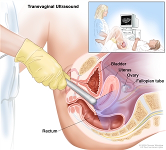
Transvaginal ultrasound. An ultrasound probe connected to a computer is inserted into the vagina and is gently moved to show different organs. The probe bounces sound waves off internal organs and tissues to make echoes that form a sonogram (computer picture).
- CT scan (CAT scan): A procedure that makes a series of detailed pictures of areas inside the body, such as the abdomen and pelvis, taken from different angles. The pictures are made by a computer linked to an x-ray machine. A dye may be injected into a vein or swallowed to help the organs or tissues to show up more clearly. This procedure is also called computed tomography, computerized tomography, or computerized axial tomography.
- Cystoscopy: A procedure to look inside the bladder and urethra to check for abnormal areas. A cystoscope is inserted through the urethra into the bladder. A cystoscope is a thin, tube-like instrument with a light and a lens for viewing. It may also have a tool to remove tissue samples, which are checked under a microscope for signs of cancer.
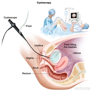
Cystoscopy. A cystoscope (a thin, tube-like instrument with a light and a lens for viewing) is inserted through the urethra into the bladder. Fluid is used to fill the bladder. The doctor looks at an image of the inner wall of the bladder on a computer monitor to check for abnormal areas.
Uterine sarcoma may be diagnosed, staged, and treated in the same surgery.
Surgery is used to diagnose, stage, and treat uterine sarcoma. During this surgery, the doctor removes as much of the cancer as possible. The following procedures may be used to diagnose, stage, and treat uterine sarcoma:
- Laparotomy: A surgical procedure in which an incision (cut) is made in the wall of the abdomen to check the inside of the abdomen for signs of disease. The size of the incision depends on the reason the laparotomy is being done. Sometimes organs are removed or tissue samples are taken and checked under a microscope for signs of disease.
- Abdominal and pelvic washings: A procedure in which a saline solution is placed into the abdominal and pelvic body cavities. After a short time, the fluid is removed and viewed under a microscope to check for cancer cells.
- Total abdominal hysterectomy: A surgical procedure to remove the uterus and cervix through a large incision (cut) in the abdomen.
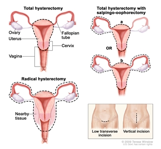
Hysterectomy. The uterus is surgically removed with or without other organs or tissues. In a total hysterectomy, the uterus and cervix are removed. In a total hysterectomy with salpingo-oophorectomy, (a) the uterus plus one (unilateral) ovary and fallopian tube are removed; or (b) the uterus plus both (bilateral) ovaries and fallopian tubes are removed. In a radical hysterectomy, the uterus, cervix, both ovaries, both fallopian tubes, and nearby tissue are removed. These procedures are done using a low transverse incision or a vertical incision.
- Bilateral salpingo-oophorectomy: Surgery to remove both ovaries and both fallopian tubes.
- Lymphadenectomy: A surgical procedure in which lymph nodes are removed and checked under a microscope for signs of cancer. For a regional lymphadenectomy, some of the lymph nodes in the tumor area are removed. For a radical lymphadenectomy, most or all of the lymph nodes in the tumor area are removed. This procedure is also called lymph node dissection.
Treatment in addition to surgery may be given, as described in the Treatment Option Overview section of this summary.
There are three ways that cancer spreads in the body.
Cancer may spread from where it began to other parts of the body.
When cancer spreads to another part of the body, it is called metastasis. Cancer cells break away from where they began (the primary tumor) and travel through the lymph system or blood.
- Lymph system. The cancer gets into the lymph system, travels through the lymph vessels, and forms a tumor (metastatic tumor) in another part of the body.
- Blood. The cancer gets into the blood, travels through the blood vessels, and forms a tumor (metastatic tumor) in another part of the body.
The metastatic tumor is the same type of cancer as the primary tumor. For example, if uterine sarcoma spreads to the lung, the cancer cells in the lung are actually uterine sarcoma cells. The disease is metastatic uterine sarcoma, not lung cancer.
Cancer can spread through tissue, the lymph system, and the blood:
- Tissue. The cancer spreads from where it began by growing into nearby areas.
- Lymph system. The cancer spreads from where it began by getting into the lymph system. The cancer travels through the lymph vessels to other parts of the body.
- Blood. The cancer spreads from where it began by getting into the blood. The cancer travels through the blood vessels to other parts of the body.
The following FIGO staging system is used for leiomyosarcomas and endometrial stromal sarcomas:
Stage I
In stage I, the tumor is found in the uterus only. Stage I is divided into stages IA and IB:
- In stage IA, the tumor is 5 centimeters or smaller.
- In stage IB, the tumor is larger than 5 centimeters.

Tumor sizes are often measured in centimeters (cm) or inches. Common food items that can be used to show tumor size in cm include: a pea (1 cm), a peanut (2 cm), a grape (3 cm), a walnut (4 cm), a lime (5 cm or 2 inches), an egg (6 cm), a peach (7 cm), and a grapefruit (10 cm or 4 inches).
Stage II
In stage II, the tumor has spread beyond the uterus but has not spread beyond the pelvis. Stage II is divided into stages IIA and IIB:
- In stage IIA, the tumor has spread to the ovary, fallopian tube, or connective tissues around the uterus.
- In stage IIB, the tumor has spread to other tissues in the pelvis.
Stage III
In stage III, the tumor has spread into tissues in the abdomen. Stage III is divided into stages IIIA, IIIB, and IIIC:
- In stage IIIA, the tumor has spread to one site in the abdomen.
- In stage IIIB, the tumor has spread to more than one site in the abdomen.
- In stage IIIC, the tumor has spread to lymph nodes in the pelvis and/or around the abdominal aorta (the largest blood vessel in the abdomen).
Stage IV
Stage IV is divided into stages IVA and IVB:
- In stage IVA, the tumor has spread into the bladder and/or the rectum.
- In stage IVB, the tumor has spread to distant parts of the body.
The following FIGO staging system is used for adenosarcomas:
Stage I
In stage I, the tumor is found in the uterus only. Stage I is divided into stages IA, IB, and IC:
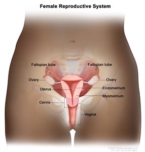
- In stage IA, the tumor is found in the endometrium or endocervix (the inner part of the cervix that forms a canal connecting the vagina to the uterus).
- In stage IB, the tumor has spread halfway or less into the myometrium (the muscular outer layer of the uterus).
- In stage IC, the tumor has spread more than halfway into the myometrium.
Stage II
In stage II, the tumor has spread outside the uterus into the pelvis. Stage II is divided into stages IIA and IIB:
- In stage IIA, the tumor has spread to the ovary, fallopian tube, or connective tissues around the uterus.
- In stage IIB, the tumor has spread to other tissues in the pelvis.
Stage III
In stage III, the tumor has spread into tissues in the abdomen. Stage III is divided into stages IIIA, IIIB, and IIIC:
- In stage IIIA, the tumor has spread to one site in the abdomen.
- In stage IIIB, the tumor has spread to more than one site in the abdomen.
- In stage IIIC, the tumor has spread to lymph nodes in the pelvis and/or around the abdominal aorta (the largest blood vessel in the abdomen).
Stage IV
Stage IV is divided into stages IVA and IVB:
- In stage IVA, the tumor has spread into the bladder and/or the rectum.
- In stage IVB, the tumor has spread to distant parts of the body.
Uterine sarcoma can recur (come back) after it has been treated.
The cancer may come back in the uterus, the pelvis, or in other parts of the body.
